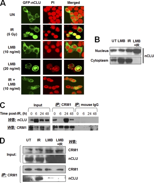
FIGURE 3.
Nuclear accumulation of hrGFP-nCLU increases after IR or LMB. A, log phase MCF-7 cells were transfected with hrGFP-nCLU or hrGFP alone. Twenty-four hours after transfection, cells were treated with IR + LMB. Forty-eight hours after transfection, cells were analyzed for hrGFP-nCLU expression with nuclei counterstained with PI. The photomicrographs shown are representative of studies performed three or more times. See Table 1 for quantification of hrGFP-nCLU nuclear localization. B, LMB enhances IR-induced nuclear accumulation of nCLU. MCF-7 cells were treated with 10 ng/ml LMB and/or 5 Gy. Cells were harvested 24 h after exposure, and the samples were separated into nuclear and cytoplasmic fractions. nCLU was detected by Western blotting using the CY-1 antibody. C, IR-dependent profiling of nCLU binding to CRM1 is shown. Whole cell extracts and immunoprecipitations (IP) were performed using the anti-CRM1 or control normal IgGs. The same CRM1 antibody was used to detect CRM1 in immunoprecipitates, and the CY-1 antibody was used to detect nCLU by Western blotting (WB). Input lanes show steady-state levels of nCLU and CRM1 proteins detected in the same whole cell extracts used for the immunoprecipitations. D, LMB (10 ng/ml) abrogates nCLU-CRM1 binding. IR-, LMB-, or IR-LMB-exposed cells were lysed, and co-immunoprecipitations were done as in C. Input indicates steady-state levels of the nCLU and CRM1 proteins detected by Western blotting in the same whole cell extracts used for the immunoprecipitations.

