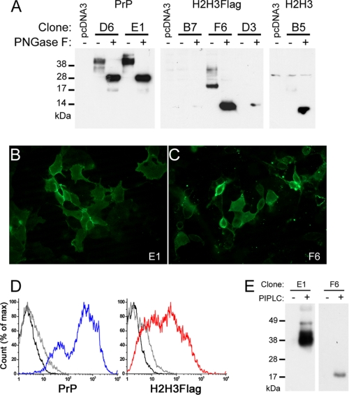
FIGURE 2.
Cell biology of selected PrP, H2H3, and H2H3Flag clones. A, study of the glycosylation of the selected clones. 10 μg of proteins from cell lysate (40 μg for B5), treated (+) or not (−) by PNGase F. D6 and E1 clones expressing PrP, B7, F6, and D3 clones expressing H2H3Flag, B5 clone expressing H2H3 were tested using 3F4, M2, and Pri-917 antibodies, respectively. B and C, fluorescence microscopy on nonpermeabilized cells. B, E1 cells displayed surface labeling of 3F4 antibody coupled with Alexa Fluor 488 secondary antibody. C, F6 cells displayed surface labeling of M2 antibody coupled with Alexa Fluor 488 secondary antibody. D, flow cytometry measurement of surface labeled PrP (blue) or H2H3Flag (red). In each plot, unlabeled control cells (black line) and isotype control labeled cells (gray line) were used. E, PrP and H2H3Flag are GPI-anchored. Western blot analysis of acetone-precipitated supernatant of PIPLC-treated (+) or untreated (−) E1 and F6 cells, using 3F4 and M2 antibodies, respectively.

