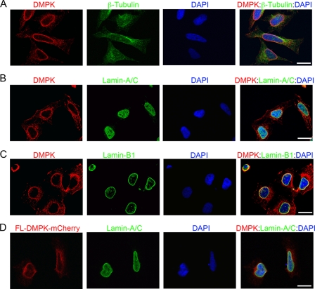
FIGURE 1.
DMPK localizes to the nuclear envelope. A–C, immunofluorescent labeling of DMPK (red) confirmed localization to the NE in HeLa cells. Scale bar = 10 μm in all panels. Staining of β-tubulin (A, green) showed cell size and shape. Colocalization with Lamin-A/C (B, green) and Lamin-B1 (C, green) confirmed DMPK at the NE. D, FL-DMPK-mCherry in HeLa cells (16-h transfection, red) colocalizes with Lamin-A/C (green). DAPI (blue) stained the nucleus in all overlay panels.

