FIGURE 2.
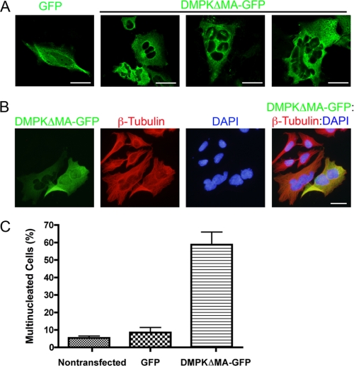
DMPK over-expression (48 h) causes nuclear fragmentation. A, overexpression of GFP (green, first panel) and DMPKΔMA-GFP (green, second through fourth panels) was compared by confocal microscopic analysis. Scale bars = 10 μm. B, overexpression of DMPKΔMA-GFP (green). β-tubulin (red) is shown together with DAPI DNA staining (blue) by standard fluorescent microscopic analysis. Scale bar = 10 μm. C, multinucleated cells were counted 48 h after transfection. Multinucleation was observed in 5.5% (non-transfected), 7% (GFP), and 58% (DMPKΔMA-GFP) of cells (n = 3 replications).

