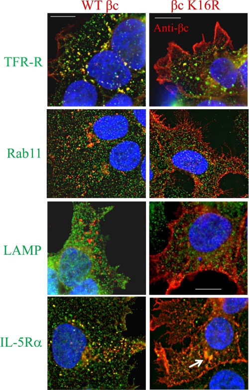
FIGURE 3.
Reduced intracellular trafficking of βc K16R at steady state. Co-localization assays were performed in unstimulated WT βc and βc K16R-expressing HEK293 cell lines by staining all cells with anti-βc antibodies (red color) and anti-transferrin receptor (top panels, green), anti-Rab11 (second panels, green), anti-LAMP-1 (third panels, green), and anti-IL-5Rα antibodies (bottom panels, green). Localization of primary antibody binding was detected by labeling with Alexa Fluor-488 (all panels, green color) or Alexa Fluor-594 (all panels, red) secondary antibodies. Images were acquired and displayed as described in the legend to Fig. 2. Yellow vesicles indicate co-localization between the two antibodies used in the assay. Note how WT βc receptors (left panels) traffic through TFR-R and Rab11-positive vesicles (yellow dots), but not LAMP-1 (lysosomal marker). Also, note the strong co-localization between WT βc and IL-5Rα in intracellular vesicles under basal conditions (bottom left). Scale bar is 10 μm.

