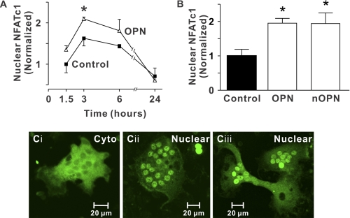
FIGURE 1.
Nuclear accumulation of NFATc1 induced by OPN. Glass coverslips were uncoated (Control) or coated with recombinant OPN (OPN) or native OPN (nOPN) as indicated. A, rabbit osteoclasts were incubated for 1.5 to 24 h. Preparations were then fixed and NFATc1 localization was assessed by immunofluorescence. Data are the proportion of osteoclasts exhibiting nuclear localization of NFATc1, expressed as a fraction of the control value at 1.5 h (mean ± S.E., n = 3 independent experiments). *, p < 0.05 compared with the control value at the same time. B, rabbit osteoclasts were plated on the indicated substrates and fixed after 3 h. The histogram shows the proportion of osteoclasts exhibiting nuclear localization of NFATc1. Data are expressed as a fraction of control and are mean ± S.E., n = 6 independent experiments. *, p < 0.05 compared with control (in this series of experiments, 19 ± 3% of osteoclasts exhibited nuclear localization of NFATc1 on uncoated (Control) surfaces). C, i, image of a osteoclast plated on an uncoated coverslip showing predominantly cytoplasmic (Cyto) NFATc1 localization. ii and iii, images of osteoclasts plated on recombinant OPN (ii) and native OPN (iii) showing NFATc1 localized predominantly in the nuclei (Nuclear). At least 2,430 osteoclasts were examined in each group.

