FIGURE 3.
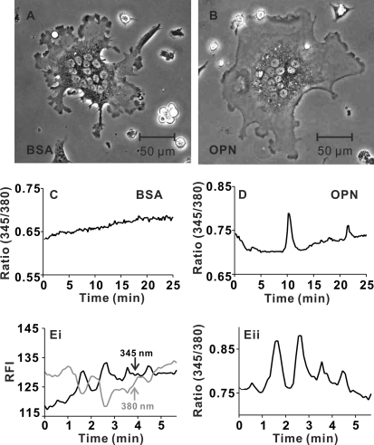
OPN enhances calcium oscillations in osteoclasts. Rat osteoclasts were plated on glass-bottomed culture dishes coated with BSA or OPN. Cells were loaded with fura-2 and bathed in supplemented M199 buffered with HEPES (25 mm) and HCO3− (4 mm). Dishes were placed in a stage warmer (35 °C) and osteoclasts were imaged using alternating excitation wavelengths of 345/380 nm. Fluorescence images were acquired every 6 s with a charge-coupled device camera. A, representative phase-contrast image of osteoclast plated on BSA. B, representative phase-contrast image of osteoclast plated on OPN. C, representative trace of the fluorescence ratio at 345/380 nm for a single osteoclast plated on BSA. In this case, oscillations in [Ca2+]i were not observed. Ratio images are available as supplemental Video S1. D, representative trace from a region of a single osteoclast plated on OPN. In this case, 2 transient elevations in [Ca2+]i were observed. Ratio images are available as supplemental Video S2. E, i, fluorescence intensities at 345 and 380 nm excitation are plotted as a function of time for another osteoclast plated on OPN. Increases in the signal at 345 nm coincided with decreases in intensity at 380 nm, consistent with authentic changes in [Ca2+]i. E, ii, fluorescence ratio for the cell illustrated in i. Data are based on responses from a total of 25–26 osteoclasts for each substrate, from 6 independent experiments.

