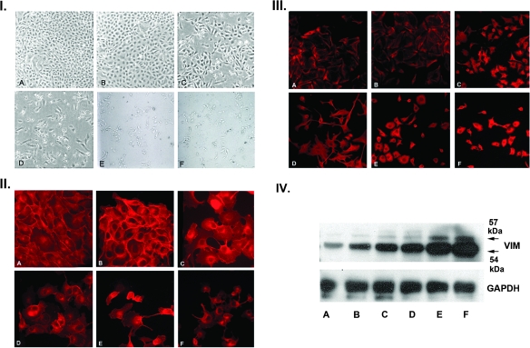
Fig. 1.
Generation and characterization of MDCK clones with a gradient of epithelial–mesenchymal transition. Panel I: MDCK cells were stably transfected with constitutively active MEK1 and selected with hygromycin as detailed in Materials and Methods. Clones were derived from picked single cells and displayed a gradient of morphologic phenotypes by phase contrast microscopy ranging from fully epithelial (B), to a mixed intermediate phenotype (C and D), to a fibroblastic mesenchymal phenotype (E and F). Panel A is a control MDCK clone transfected with pTK-hydro alone. Panel II: Immunofluorescence (IF) staining of MDCK clones for E-cadherin. E-cadherin is progressively lost from junctional complexes as a function of the extent of epithelial–mesenchymal transition and assumes a primarily cytosolic and perinuclear distribution, particularly in the most mesenchymal E and F clones. Panel III: IF staining of MDCK clones for vimentin. In the epithelial clones, vimentin is found in a subcortical distribution, but as epithelial–mesenchymal transition progresses, there is dense cytocolic and perinuclear staining with vimentin arranged in cord-like bundles (I–III, ×200). Panel IV: Western blot analysis of vimentin in the MDCK clones. There is a progressive increase in the expression of vimentin in the MDCK clones as a function of the extent of epithelial–mesenchymal transition.

