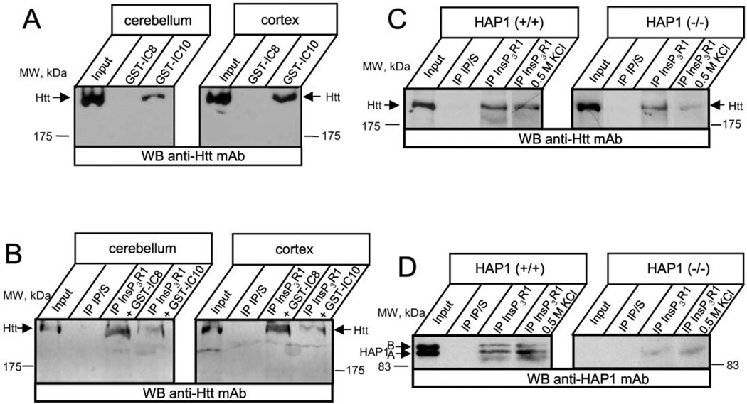
Figure 2. InsP3R1 Is Associated with Htt In Vivo.
(A) Rat cerebellar (left) or cortical (right) lysates were used in GST-IC8/IC10 pull-down experiments.
(B) Rat cerebellar (left) or cortical (right) lysates were used in coimmunoprecipitation experiments with anti-InsP3R1 polyclonal antibodies or corresponding preimmune sera (IP/S). GST-IC8 and GST-IC10 proteins (200 µg/ml final concentration) were included in the immunoprecipitation reactions as indicated. The precipitated fractions on (A) and (B) were analyzed by Western blotting with anti-Htt monoclonal antibodies.
(C and D) Cortical lysates from wild-type (left panel) or HAP1−/− (right panel) mice (at postnatal day 3) were used in immunoprecipitation experiments with the anti-InsP3R1 polyclonal antibodies or with the corresponding preimmune sera (IP/S). The precipitated proteins were analyzed by Western blotting with the anti-Htt monoclonal antibodies (C) or with the anti-HAP1 monoclonal antibodies (D). An additional 0.5 M KCl wash step was included in the immunoprecipitation protocol as indicated. The input lanes on (A)–(D) contain 1/50 of lysates used in immunoprecipitation and GST pull-down experiments.

