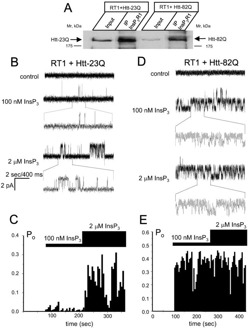
Figure 5. Full-Length Httexp Sensitizes InsP3R1 to Activation by InsP3.
(A) Microsomes from Sf9 cells coinfected with RT1 and Htt-23Q or Htt-82Q baculoviruses were solubilzed in 1% CHAPS, precipitated with anti-InsP3R1 polyclonal antibodies, and blotted with the anti-Htt monoclonal antibodies. The input lanes contain 1/10 of lysates used in the immunoprecipitation experiments.
(B) Activity of InsP3R1 coexpressed in Sf9 cells with Htt-23Q and reconstituted into planar lipid bilayers. Responses to application of 100 nM InsP3 and 2 µM InsP3 are shown. Each current trace corresponds to 10 s (2 s for expanded traces) of current recording from the same experiment.
(C) The average InsP3R1 open probability (Po) was calculated for a 5 s window of time and plotted for the duration of an experiment. The times of 100 nM and 2 µM InsP3 additions to the bilayer are shown above the Po plot. Data from the same experiment are shown on (B) and (C). Similar results were obtained in four independent experiments.
(D and E) Same as (B) and (C) for InsP3R1 coexpressed in Sf9 cells with Htt-82Q. Similar results were obtained in six independent experiments.

