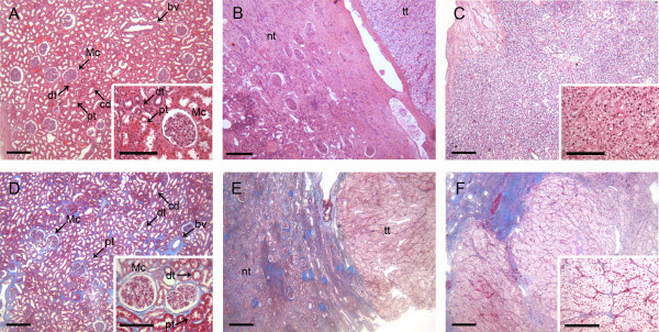
Figure 1.
Representative images of hematoxylin & eosin (HE) and azan stained human kidney tissue sections. A-C, H&E-stained kidney sections. D-F, Azan-stained kidney sections. A and D show the renal cortex of normal kidney tissue. B and E present kidney sections with areas of healthy tissue and clear cell renal cell carcinoma (intermediate kidney tissue). C and F show sections of CCRCC. Mc: Malpighian corpuscle, dt: distal tubule, pt: proximal tubule, cd: collecting duct, bv: blood vessel, tt: tumor tissue, nt: normal tissue. Scale bars: 300 μm, scale bars inset: 150 μm.

