Figure 3.
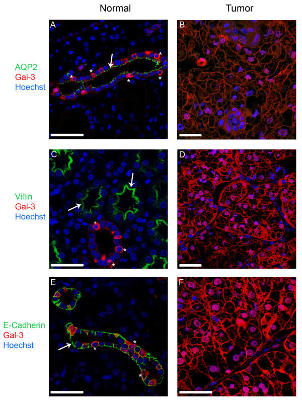
Confocal fluorescence images showing the distribution of galectin-3 and different polarity markers in normal kidney and tissue from clear cell renal cell carcinoma. All sections were immunostained against apical aquaporin-2 (AQP-2) and villin or basolateral E-cadherin. In all fluorescence images the polarity markers are indicated in green, galectin-3 is depicted in red and the nuclei are stained with Hoechst 33342 (blue). In normal kidney sections aquaporin-2 is concentrated on the apical domain of epithelial cells of the collecting duct, whereas villin is part of the brush border of the proximal tubule. E-cadherin can be detected in cells of the distal tubule and the collecting duct. Arrows mark the apical localization of AQP-2 and villin (A, C) or the basolateral localization of E-cadherin (E). In all tissue sections of the tumor the expression of the polarity markers is reduced or completely lost. In normal kidney areas, galectin-3 is found in the collecting duct as well as in the distal tubule, but not in the proximal tubule. Stars depict single cells, in which galectin-3 is expressed. Scale bars: 25 μm.

