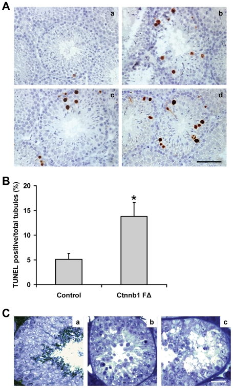
Figure 3. Increased germ cell apoptosis in Ctnnb1 FΔ mice testes.
(A) TUNEL analysis of apoptotic germ cells in control (panel a) and Ctnnb1 FΔ (panels b-d) mice testes sections. Scale bar, 50 µm. (B) Number of TUNEL-positive cells per seminiferous tubule (n = 6, *p<0.05). (C) Light micrographs of epox-embedded and toluidine blue-stained testicular sections from control and Ctnnb1 FΔ mice. Tubular profiles from a control mouse testis showing Sertoli cells and germ cells at various phases of development that support normal spermatogenesis (panel a). Tubular profiles from a Ctnnb1 FΔ mouse showing epithelial vacuolization and complete loss of elongated spermatids (panels b and c). Scale bar, 50 µm.

