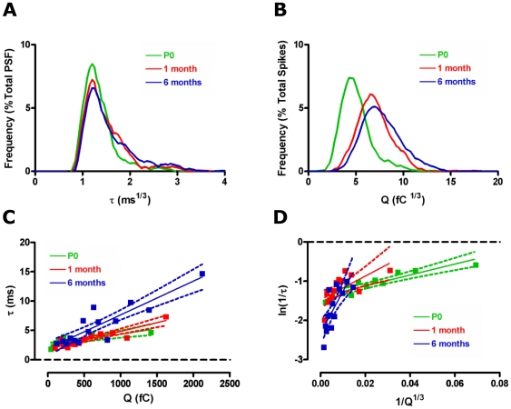
Figure 6. Aging differentially affects membrane bending properties during exocytosis.
Frequency distribution of the cube root of pre-spike foot (PSF) signal duration (A) and spike area (B) illustrates the different ages at which these change. Correlations between PSF signal duration, τ, and spike area, Q (C) shows a linear relationship at all ages (P0: R2 = 0.64, p<0.01, 1 month: R2 = 0.82, p<0.0001, 6 months: R2 = 0.79, p<0.0001). Transforms of this data to ln(1/τ) vs 1/Q1/3 (D) also provides linear relationships at all ages (P0: R2 = 0.81, p<0.0001, 1 month: R2 = 0.44, p<0.05, 6 months: R2 = 0.59, p<0.01). The slope of this plot, representing membrane curvature [1], increases significantly with age. Green – P0, red – 1 month, blue – 6 months.

