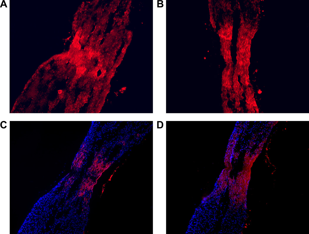
Figure 2.
Auto fluorescence micrographs from a 14 day severed tendon explant that was incubated with red fluorescent fibril segments for 4 days. Panels A and B show the accumulation of auto-fluorescence within the wound gap between organ cultured severed tendon explants. Panel C and D show DAPI blue stained fluorescence tendon fibroblast nuclei along with red auto-fluorescent fibril segments, which have amassed at the severed tendon wound gap. Note in panel D the absence of stained nuclei at the center of the wound gap, suggesting the accumulation of fibril segments on growing collagen fibers can be independent of direct association with tendon fibroblasts.

