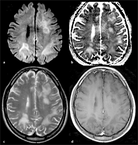
Fig. 1.
Brain MRI showing bilateral multifocal white matter lesions. The lesions contain high signal-intense rims on diffusion-weighted images (a). With low signal intensity on an afferent diffusion coefficient map (b). The findings are consistent with demyelinating plaque. c T2-weighted images display hyperintense lesions with surrounding edema. d Gadolinium-enhanced T1-weighted image does not reveal an enhanced lesion area.

