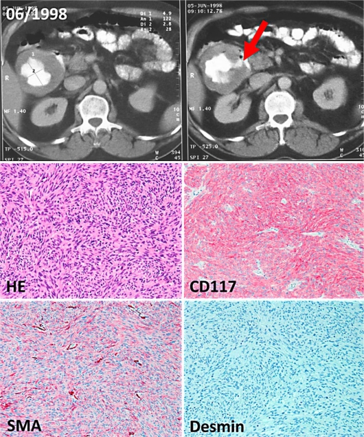
Fig. 1.
Upper panel: CT scan of the abdomen showing a cystic abdominal mass. The necrotic tumor of the duodenum is filled with contrast medium, the perforation to the duodenum is indicated with an arrow. Lower panel: Representative paraffin sections of the duodenal GIST (original magnification 200×). The sections show a spindle cell (HE staining), ckit-positive (CD117), smooth muscle actin (SMA)-positive and desmin-negative mesenchymal tumor.

