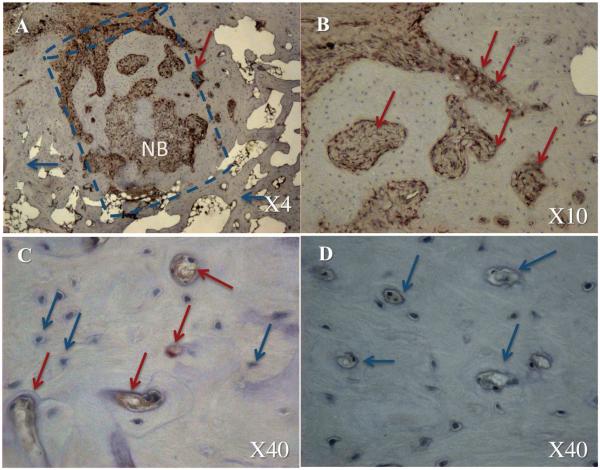
Figure 7. Stem cell contribution to the bone void repair: immunohistochemical analysis.
The dashed line shows the estimated borders of the defect. NB – new bone formation. The blue arrows indicate a native host cell and the red arrows indicate donor porcine cells. The defect repaired with gene-modified stem cells is shown in panels A–C, whereas defect treated with FG alone is shown in panel D.

