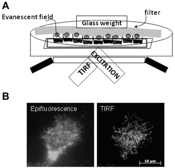
Figure 1.

Total internal reflection fluorescence microscopy (TIR-FM) can be used to study events at the apical membrane. (A) Schematic of apical TIR-FM setup. OKP cells transfected with a fluorescent-tagged protein of interest are grown to confluence on a filter. The filter is cut out and placed upside down in a dish with a coverslip bottom. A glass weight is used to keep the filter in place. Incident laser light is totally internally reflected to produce an evanescent field that only illuminates the brush border microvilli. (B) Comparison between an opossum kidney cell transfected with green fluorescent protein-tagged Npt2a and imaged with conventional epifluorescence (left) versus apical TIR-FM (right). Note that, with apical TIR-FM, the resolution of Npt2a within the microvilli of the brush border membrane is much better than that of conventional epifluorescence. Figure has been adapted from Blaine et al. Am J Physiol Cell Physiol. 2009;297:C1339-C1346.
