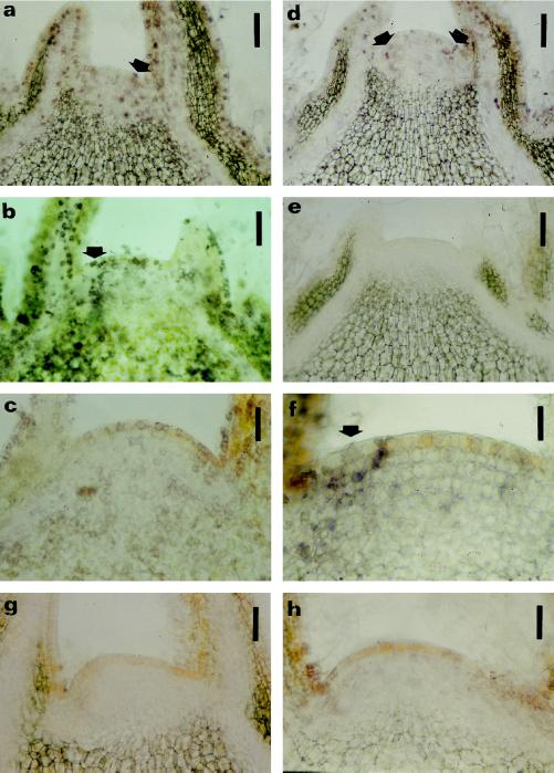
Figure 3.
Immunolocalization of cytokinin bases within vegetative shoot apices. The location of cytokinin bases in longitudinal sections was determined using immunohistochemistry. Nitroblue tetrazolium/5-bromo-4-chloro-3-indolyl phosphate was used as chromogenic substrate for the AP, resulting in a purple reaction product. Immunolocalization of zeatin (a and d), DHZ (b and e), and IP (c and f) in vegetative shoot apices at developmental stages I1 (a, b, and c) and I2 (d, e, and f) (Fig. 1). Purple staining is present for zeatin (a and d) in the nucleus and cytoplasm, whereas for DHZ (b and e) and IP (c and f) a more perinuclear purple staining is visible. Arrows in a, b, d, and f point to staining in the lateral zones of the meristems. g and h, Sections (from stage I1, see Fig. 1) incubated with anti-zeatin (g) and anti-DHZ (h) antibodies saturated with zeatin and DHZ as a control, respectively. Bars = 90 μm (a, b, d, e, g, and h); 45 μm (c); and 22 μm (f).

