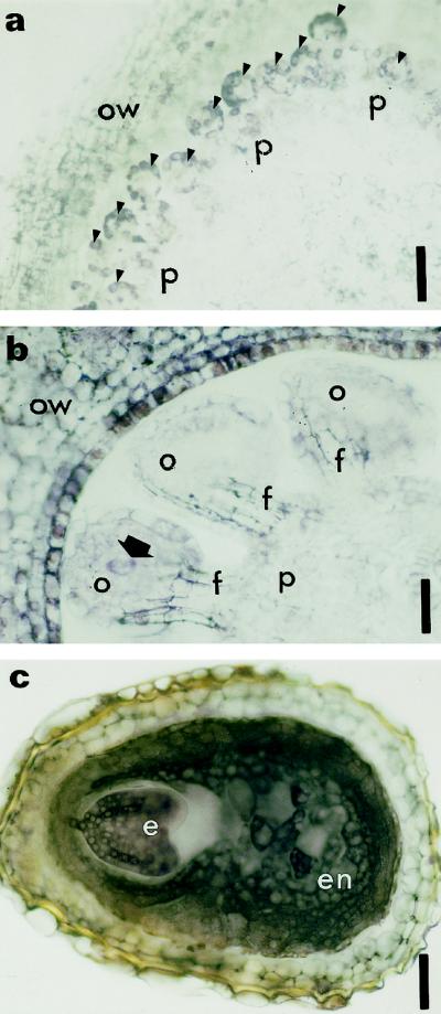
Figure 6.
Immunolocalization of zeatin at two stages of ovule formation and at the end of seed formation. Cross-sections of ovule primordia arising from the placenta (a, arrowheads) and developing ovule (b, arrow indicates the archesporial cell in a developing ovule). Purple staining resulting from labeling of zeatin was detected in ovule primordia (a, arrowheads), the ovary wall (a and b), and the developing ovules (b). Archesporial cells are also significantly labeled (b). c, Cross-section of a seed immunolabeled with anti-zeatin antibody. The cytoplasm of embryo and endosperm are heavily stained. e, Embryo; en, endosperm; f, funiculus; o, ovule; ow, ovary wall; p, placenta. Bars = 180 μm (a) and 90 μm (b and c).

