
Figure 3.
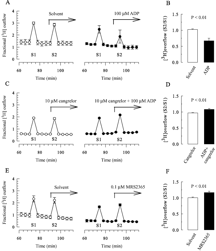
Modulation of [3H]-noradrenaline release from SCG neurons by ADP, MRS 2365 and cangrelor. SCG cell cultures were labelled with [3H]-noradrenaline and superfused. Subsequent to a 60 min washout period, 4 min fractions of superfusate were collected, and tritium overflow was evoked by electrical field stimulation as shown in Figure 1A. (A, C and E) Exemplary time courses of fractional 3H outflow as a percentage of the total radioactivity in the cells (n = 3); 100 µM ADP, 10 µM cangrelor, 0.1 µM MRS 2365 or the appropriate solvent were present from minute 88 onwards as indicated by the arrows. (B) S2/S1 ratios obtained in the presence of either solvent or 100 µM ADP (n = 11). (D) S2/S1 ratios obtained in the presence of either 10 µM cangrelor or 10 µM cangrelor plus 100 µM ADP (n = 6). (F) S2/S1 ratios obtained in the presence of either solvent or 0.1 µM MRS 2365 (n = 12); the P-values for the statistical significances of differences (Student's unpaired t-test) are indicated above the bars.