
Figure 4.
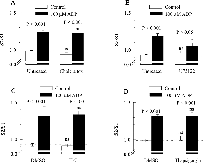
The enhancement of [3H]-noradrenaline release from rat SCG neurons by ADP involves phospholipase C, but not Gs-proteins, PKC or Ca2+-ATPase. All SCG cell cultures were treated with pertussis toxin (PTX; 100 ng·mL−1 for 24 h). In addition, some cultures were treated with cholera toxin or remained otherwise untreated. Thereafter, the cultures were labelled with [3H]-noradrenaline in the absence (untreated) or presence of 3 µM U73122 and were then superfused. Subsequent to a 60 min washout period, 4 min fractions of superfusate were collected, tritium overflow was evoked by electrical field stimulation, and ADP was applied as shown in Figure 1A. When appropriate, 10 µM H-7, 0.3 µm thapsigargin, or 0.1% DMSO were present from minute 50 of superfusion onwards. (A) The S2/S1 ratios of tritium overflow in the absence (control) or presence of 100 µM ADP in either untreated or cholera toxin-treated neurons (n = 10–12). (B) The S2/S1 ratios of tritium overflow in the absence or presence of 100 µM ADP in either untreated or U73122-treated neurons (n = 9). (C) The S2/S1 ratios of tritium overflow in the absence (control) or presence of 100 µM ADP applied either in a solution containing DMSO, or in a solution containing H-7 (n = 9). (D) The S2/S1 ratios of tritium overflow in the absence (control) or presence of 100 µM ADP applied either in a solution containing DMSO or in a solution containing thapsigargin (n = 9). P-values for the significance of differences between the results obtained in the absence and presence of ADP are indicated above the bars; * indicates a significant difference versus the corresponding result obtained in untreated cultures at P < 0.05; ns indicates no significant difference versus results obtained in either untreated cultures (A and B) or in the presence of DMSO (C and D).