Figure 3a.

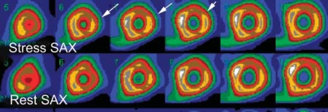
The size or extent of the myocardial perfusion in three different patients. Selected short-axis images (SAX) at stress and rest. Small size (less than 10% of the LV myocardium) reversible anterolateral wall perfusion defect (arrows).

The size or extent of the myocardial perfusion in three different patients. Selected short-axis images (SAX) at stress and rest. Small size (less than 10% of the LV myocardium) reversible anterolateral wall perfusion defect (arrows).