Figure 4.

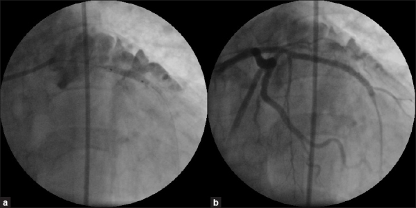
(a) Fluoroscopy showing deployment of a covered stent within a previous stent at the perforation site. (b) Left coronary angiogram post- deployment of a covered stent showing complete sealing of left anterior descending artery perforation

(a) Fluoroscopy showing deployment of a covered stent within a previous stent at the perforation site. (b) Left coronary angiogram post- deployment of a covered stent showing complete sealing of left anterior descending artery perforation