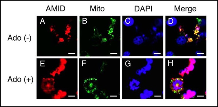
Fig. 3.
Immunofluorescent cytochemistry. HuH-7 cells expressing HA-AMID were untreated [Ado (+)] and treated with adenosine (10 mM) for 4 h [Ado (+)]. Then, cells were reacted with an anti-HA antibody for AMID (red), MitoTracker Green FM for the mitochondria (Mito)(green) and DAPI for the nucleus (blue). Similar results were obtained with 4 independent experiments. Bars, 10 μm.

