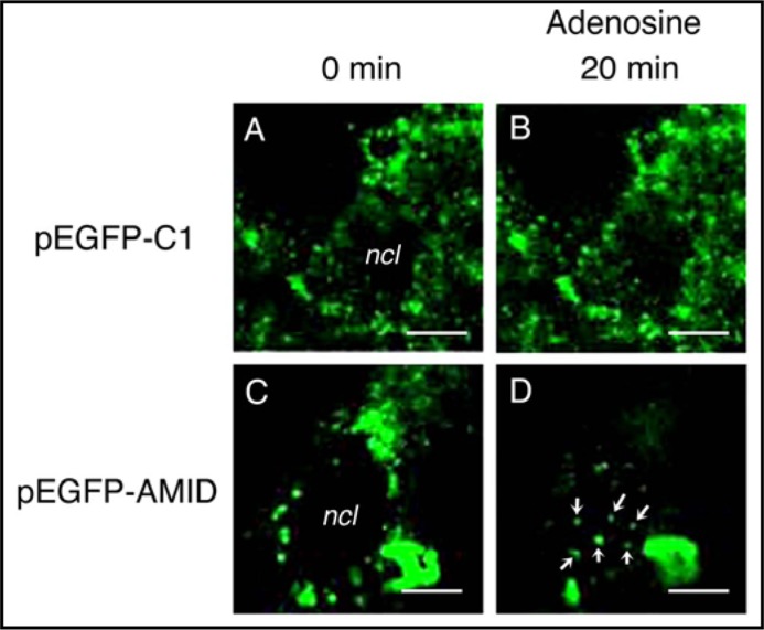
Fig. 4.
Time-laps monitoring for intracellular AMID mobilizations in living HuH-7 cells. Intracellular GFP signals were monitored in cells expressing GFP alone (pEGFP-Cl)(A, B) or GFP-AMID (pEGFP-AMID)(C, D) every 5 min before and after treatment with adenosine (10 mM). Note accumulation of GFP signals in the nucleus (ncl) 20 min after adenosine treatment (arrows) for cells expressing GFP-AMID. Similar results were obtained with 4 independent experiments. Bars, 10 um.

