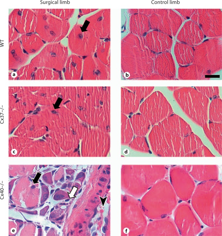
Fig. 3.
Representative images of cross-sections of the gastrocnemius muscle stained with hematoxylin and eosin and harvested from the surgical and control limbs of WT (a, b), Cx37–/– (c, d) and Cx40–/– (e, f) animals at day 14 after FSAVPR (scale bar = 20 μm). Centrally located muscle fiber nuclei (black arrows) were observed in tissue obtained from the surgical limbs of all genotypes, indicating muscle fiber damage and regeneration. Additionally, tissue from the surgical limbs of Cx40–/– animals exhibited substantial disorganization, hemorrhage (arrowhead), and interfiber eosinophilic staining (white arrow), indicative of the increased severity of ischemic damage in these animals.

