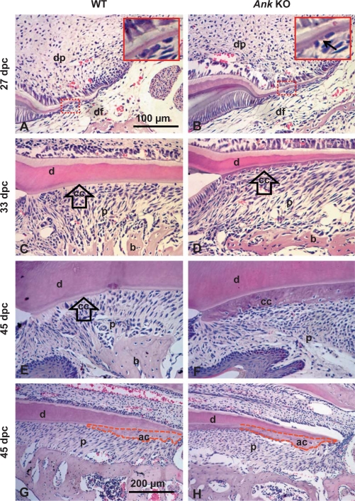
Fig. 2.
Loss of ANK causes cementum hyperplasia. 27 dpc: root formation was underway by 27 dpc, with cervical cementum barely visible in WT at this stage (A) while Ank KO mouse molars (B) exhibited regions of abnormally thick cementum matrix (indicated by black arrow in inset). 33 dpc: by 33 dpc, root development advanced apically, root dentin (d) thickened, and acellular cervical cementum covered dentin in WT (C). In the Ank KO mouse (D), cervical cementum (cc) was several-fold expanded versus WT and cells were partially embedded in the matrix. 45 dpc: in WT mice at 45 dpc, the first molar root was principally complete with both acellular (cervical) (E) and cellular (apical) (G) cementum (ac). In the Ank KO mouse molar, the widened cervical cementum (F) included numerous cell inclusions (cementocytes). PDL space (p) was maintained. Conversely, KO apical cementum (H) was not different from WT. Apical cementum is marked by a dotted red outline. All images are from lingual side of first mandibular molar. Scale bars represent 100 μm (A–F) or 200 μm (G, F). dp = Dental pulp; DF = dental follicle; b = bone.

