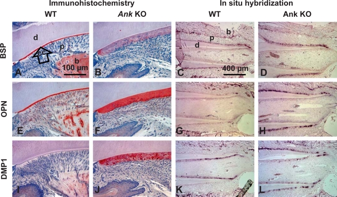
Fig. 5.
Loss of ANK alters cementoblast gene expression and cervical cementum extracellular matrix composition. Histological sections from 45 dpc mice were used for IHC, while 33 dpc tissues were used for ISH. BSP: in WT (A), BSP protein staining was strong in bone (b) and defined the entire acellular cementum (cc). Ank KO cervical cementum (B) showed diffuse and limited BSP staining. ISH revealed Bsp mRNA expression in cementoblasts and osteoblasts in WT tissues (C). Ank KO cementoblasts expressed similar levels of Bsp mRNA (D). OPN: in WT (E), OPN was observed at bone cement lines, in PDL (p), and strongly labeled acellular cementum. OPN localization in KO (F) was similar to WT, with cervical cementum exhibiting strong and even OPN labeling. Opn mRNA expression was localized to bone-lining osteoblasts and some cementoblasts, especially those apically located (G). In Ank KO, Opn mRNA was intensely expressed by cementoblasts lining the entire (cervical) cementum (H). DMP1: DMP1 in WT (I) was localized to bone around osteocytes, but not acellular cementum. In Ank KO (J), DMP1 was strongly localized to cervical cementum, particularly to perilacunar spaces around cells. By ISH, Dmp1 mRNA was identified in WT odontoblasts and osteocytes (K). Dmp1 message was clearly increased in cementoblasts/cementocytes along the molar root of the Ank KO mouse, but levels were not different in odontoblasts, osteoblasts and osteocytes of Ank KO versus WT (L). For IHC, lingual aspect of mandibular first molar is featured; see online supplementary figure 3 for overall protein distributions in the dentoalveolar complex. Scale bars represent 100 μm in A, B, E, F, I and J,and 400 μm in C, D, G, H, K and L. d = Dentin.

