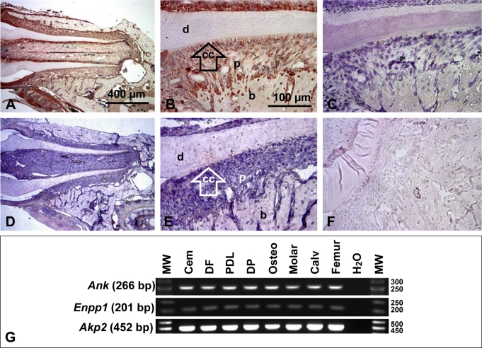
Fig. 6.
ANK is widely expressed during tooth root development. A, B In 33 dpc WT molars, ANK protein staining was observed in cells and tissues of the developing dentoalveolar complex, e.g. ameloblasts, odontoblasts, PDL, cementoblasts, osteoblasts and osteocytes. These representative images were from use of ANK-3 antibody; results with additional ANK antibodies are shown in online supplementary figure 5. While ANK staining was widespread in tooth tissues, it was not matched in negative controls (C) or Ank KO sections (online suppl. fig. 5). (D, E). In situ hybridization for Ank mRNA in 33 dpc WT tissues confirmed widespread immunohistochemical results whereas sense negative controls were not stained (F). Scale bars represent 400 μm in A and D, and 100 μm in B, C, E and F. G Tooth-derived cells and tissues express Ank, Enpp1, and Akp2: Cementoblasts (Cem), dental follicle (DF), PDL cells, primary dental pulp (DP) cells, pre-osteoblasts (Osteo), and whole mouse molar, calvaria (Calv), and femur RNA preparations.

