Figure 3.
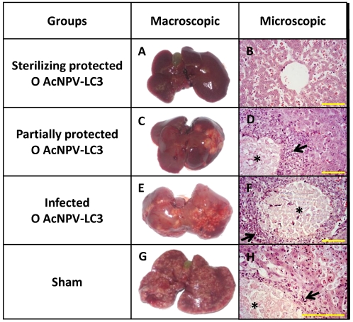
ALA development and hepatic damage analysis in AcNPV-LC3 immunized hamsters after challenge with E. histolytica. Representative pictures of the three levels of ALA development observed in hamsters of the O AcNPV-LC3 group, completely protected (sterilizing protection, A), partially protected (C) and non-protected (E). For comparative purposes, one representative liver from an animal of the sham group is shown in G. Hematoxylin-eosin stained section from each liver is shown in B, D, F and H, respectively. Healthy liver tissue with no trophozoites was observed in the completely protected hamsters (A and B), in contrast with local (partially protected; C and D) and spread (unprotected; E to H) abscesses, where necrotic focus with multiple trophozoites (*) surrounded by inflammatory infiltrates (arrow), are usually observed. Yellow bars: 100 µm.

