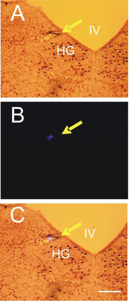
Figure 4. Histological identification of the microinjection sites in the medulla oblongata.
The injection sites of l-Glu identified by the deposit of fluorescent microbeads which were injected at the same sites of l-Glu microinjection at the end of the experiment. A: Brain stem section stained with neutral red. B: Computer image of fluorescent microbeads. C: Image of brain stem section overlapped with computer image of fluorescent microbeads. IV, fourth ventricle; HG, hypoglossal nucleus. Arrow indicates site of microinjection. Calibration bar: 500 µm.

