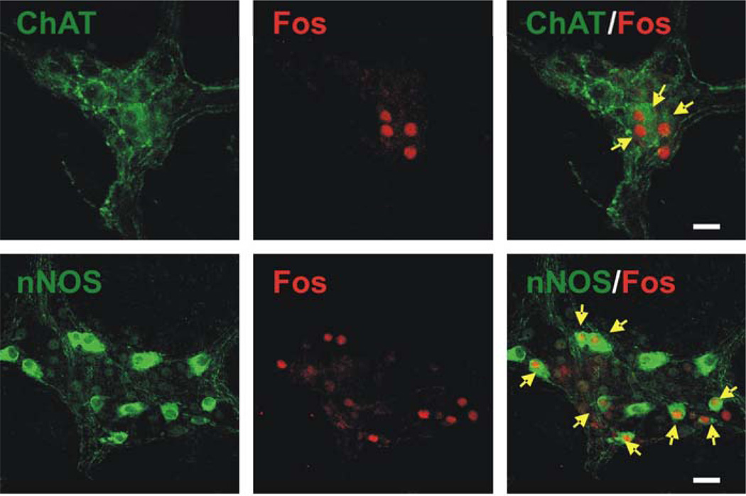
Figure 7. Immunohistochemical demonstration of double labeling for c-Fos and choline acetyltransferase (ChAT) or nitric oxide synthase (nNOS) in intragastric myenteric ganglionic neurons in whole mount preparation.
Stimulation of DMV neurons in the rostral part of the nucleus (upper panel) induced Fos immunoreactivity (Cy3, red) in intragastric myenteric neurons that mainly contain ChAT. In contrast, stimulation of DMV neurons in the caudal part of the nucleus (lower panel) induced FOS immunoreactivity in intragastric myenteric neurons that mainly contain nNOS. Arrows indicate c-Fos–positive ChAT- or nNOS containing neurons. Calibration bar: 50 µm.

