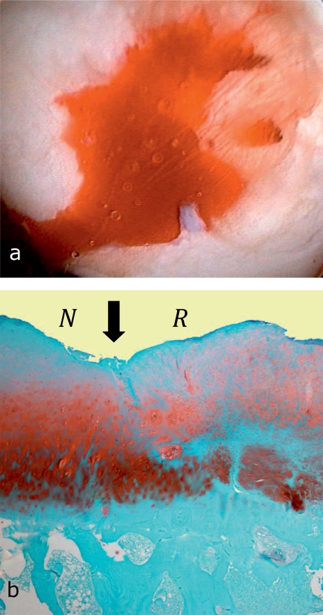
Figure 2.
Arthroscopic view of microfracture surgery and histological finding after microfracture surgery
Arthroscopic view in microfracture surgery: the cartilage defect was carefully débrided, the calcified cartilage meticulously removed, and stable walls created around the edges. After multiple microfractures have been applied with a microfracture chisel, threads of blood appear from the channels created by the microfracture—at low arthroscopic pump pressure—and form the basis for the subsequent repair tissue.
In the histology image after microfracture (Safranin O—fast green the original cartilage(left, N) shows signs of degeneration; the repair tissue (right, R) is characterized by round cells, characteristic for chondrocytes, and a proteoglycan-containing extracellular matrix. Arrow: integration of the repair tissue with the neighboring original hyaline cartilage.

