Figure 3.
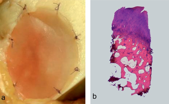
Intraoperative findings in matrix-associated autologous chondrocyte implantation (ACI) and osteochondral biopsy after matrix-associated ACI.
Intraoperative finding in matrix-associated autologous chondrocyte implantation. The chondrocytes are distributed in a biologically degradable matrix that inserted into the cartilage defect and fixated with resorbable sutures (size USP 6–0).
Osteochondral biopsy (hematoxylin and eosin) from a 39-year-old patient after ACI (primary diagnosis: traumatic cartilage injury, medial femoral condyle, 4.3 cm2). Clinically, the patient is asymptomtomatic. Histology shows a fibrocartilaginous repair tissue with early signs of degeneration of the superficial cartilage layer; hyaline cartilage is visible in the deeper layers of the repair tissue.

