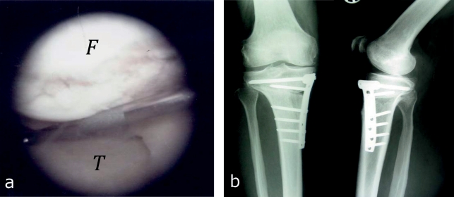
Figure 4.
Arthroscopic view of medial varus gonarthrosis and radiograph after open-wedge high tibial osteotomy
Arthroscopic view, medial compartment with osteoarthritic cartilage defects in the area of the medial femoral condyle (F) and the medial tibial plateau (T).
Radiograph after open-wedge high tibial osteotomy in medial valgus gonarthrosis. The osteotomy was performed in a biplanar technique: horizontal osteotomy of the posterior two thirds of the tibia is performed first; this is followed by an osteotomy with a 110 degrees cut behind the tuberosity parallel to the ventral tibial shaft. The result of the correction is fixated with a plate fixatior.
.

