Fig. 4.
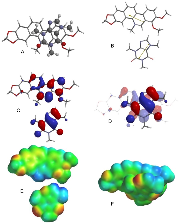
Lowest-energy orientation of the π–π complex between chelerythrine and caffeine. (A) Face-to face orientation of caffeine (ball and spoke model) in its lowest-energy orientation with chelerythrine (tube model). (B) Molecular dipoles of chelerythrine (top) and caffeine (bottom). (C) LUMO of chelerythrine (top) and HOMO of caffeine (bottom). (D) Frontier molecular orbital overlap of caffeine with chelerythrine in the lowest-energy orientation. (E) Electrostatic potential maps of chelerythrine (top) and caffeine (bottom). (F) Electrostatic potential map of the lowest-energy π–π complex between chelerythrine and caffeine.

