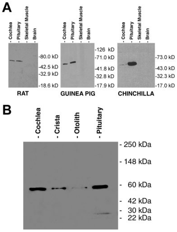
Fig. 1.
A: Western blots of rat, guinea pig, and chinchilla immunoreactivity in cochlea, pituitary, skeletal muscle, and whole-brain homogenates. Thirty micrograms of tissue protein was loaded for each lane. Cochlear samples were not microdissected, but rather tissue was homogenized with some surrounding bone present. B: Western blot demonstrating the variation in levels of reactivity among two cochleas, six cristae, four otolith organs, and a pituitary from one chinchilla. Vestibular and cochlear end organs were microdissected in this case, thus minimizing the amount of bone present. In order not to overload the lanes, 22 µg of cochlear protein (one-tenth of one cochlea), 72 µg protein from six microdissected cristae (all the cristae present in one animal), 60 µg of four microdissected otolith organs (one animal’s otolith organs), and 30 µg of pituitary protein (one-fifth of one pituitary) were used.

