Figure 1.
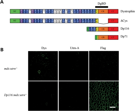
Expression of dystrophin, utrophin and Dp116 in transgenic mice. (A) Domain structure of dystrophin and selected isoforms tested as transgenes in mdx:utrn−/− mice. NT, NH2-terminal actin-binding domain; H , hinge; R, spectrin-like repeat; W, WW domain; CR, cysteine-rich domain; CT, carboxy-terminal domain, DgBD, dystroglycan-binding domain; F, Flag epitope tag. Both Dp71 and Dp116 have unique NH2-terminal peptides, shown in green. The Dp116 protein expressed in this study has a Flag epitope tag incorporated upstream of the unique peptide. Basic spectrin-like repeats that contribute to the central rod actin-binding domain are shown in white. (B) Immunofluorescent staining of quadriceps muscles for full-length dystrophin, utrophin-A and Dp116. Dystrophin (Dys) is only detected in rare, revertant myofibers in mdx:utrn−/− mice. Utrophin-A (Utrn-A) is not detected in any of the mice analyzed. Flag epitope-tagged Dp116 is localized to the sarcolemma in transgenic Dp116:mdx:utrn−/− myofibers. Scale bar: 100 μm.

