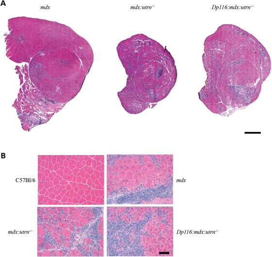
Figure 3.
Histology of quadriceps muscles from 8-week-old transgenic and control mice. (A) Entire cross-sections from the mid-belly of quadriceps muscles indicate extensive degeneration in both mdx:utrn−/− and transgenic Dp116:mdx:utrn−/− muscles compared with mdx. Scale bar: 1 mm. (B) High power (20×) fields of the muscles shown above along with wild-type control. Pathology of mdx and mdx:utrn−/− skeletal muscles is qualitatively similar at this age, with areas of extensive necrosis, inflammatory infiltrate and large numbers of myofiber containing centrally located nuclei. Similar pathologic changes are seen in the Dp116 transgenic muscles. Scale bar: 50 μm.

