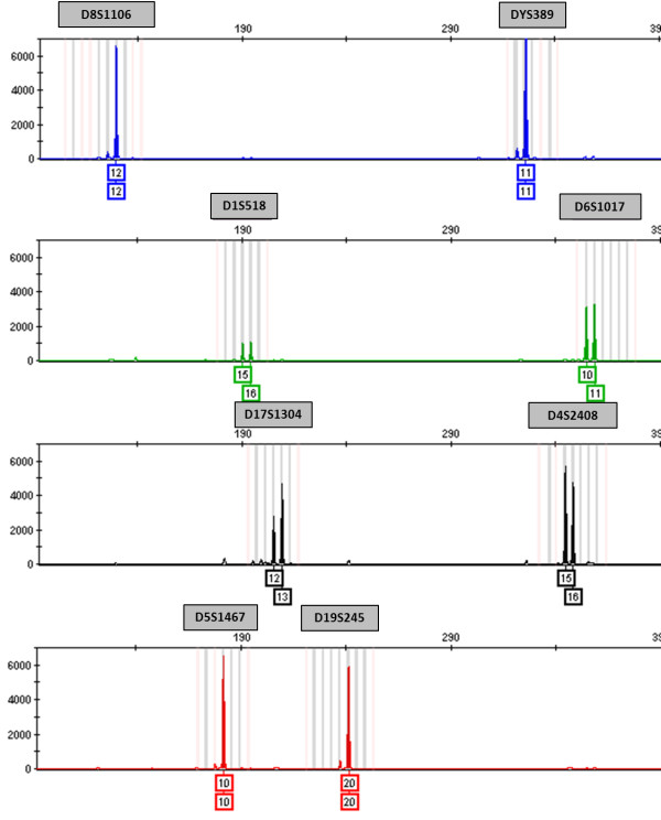
Figure 1.
Genetic profile of the Vero cell line using the vervet multiplex assay and bins and panels developed in GeneMapper ID-X. The blue, green, black, and red peaks in the electropherogram correspond to the fluorescently labeled forward primers for that STR marker (FAM, VIC, NED, and PET, respectively). Relative fluorescent units (RFUs) are depicted on the y-axis and fragment length on the x-axis. The number(s) below each peak represent the number of repeats at that locus. Vero peaks appear in the shaded bins and panels designated for each STR marker. The bins represent individual alleles for a specific marker.

