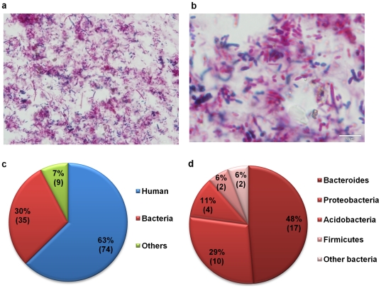
Figure 2. Cellular and protein composition at the MLI.
Upper panel: Cytology analysis of the cell pellet obtained from each mucosal lavage sample using gram staining. a.100× b. 500×. Lower panel: Distribution of proteins with different origins identified from the mucosal lavage sample using shotgun proteomic analysis. c. Composition of proteins from all species as identified by tandem MS. Other origin includes phage and amoebozoa. d. Composition of bacterial proteins. Other bacterial origin includes Chlorobi and Cyanobacteria.

