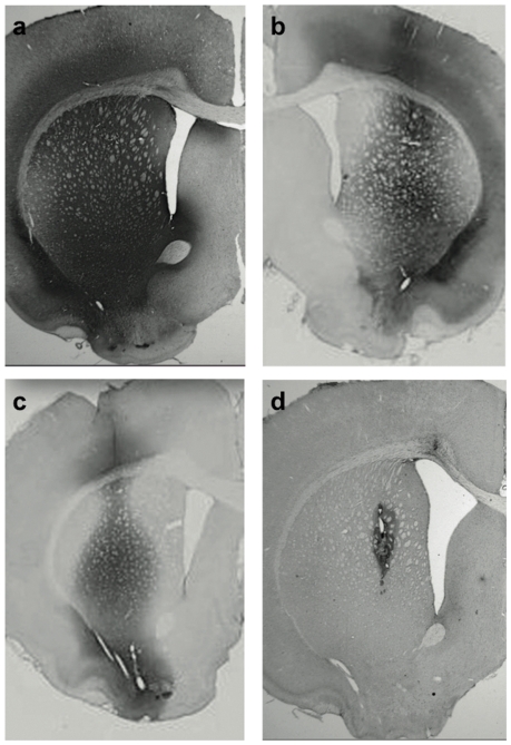
Figure 3. Immunostaining for GDNF expressed in rat striatum from constitutive AAV2-GDNF (a-c) and rapamycin-regulated AAV2-GDNF (d).
The constitutive vector was infused at 3 different doses: 16.5×10¹⁰ vg (a), 9.07×10¹⁰ vg (b), and 1.65×10¹⁰ vg (c). The rats were euthanized 4 months after transduction. Robust, widespread signal (proportional to vector dose) confirmed continuous GDNF expression within the injected hemisphere. Much of the signal represents GDNF secreted into the striatal parenchyma. The dose of AAV2-regGDNF was 4.12×10¹⁰ vg and rats were euthanized 4 days after 3-day Rapamycin regimen (3×10 mg/kg). In contrast to the constitutive vector, the AAV2-regGDNF revealed only focal expression of GDNF localized mainly around the cannula track (d). Since this vector induces GDNF secretion only in the presence of rapamycin, the GDNF signal was limited and did not extend beyond the injected striatum (no GDNF accrual upon a single rapamycin cycle).

