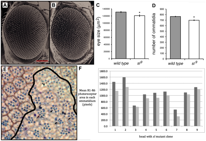
Figure 3. Sl affects eye size. Images in (A, B) are SEMs of control (A; Canton S) and (B) sl9 homozygote female flies.
Notice mild roughness of sl9 mutant eye; red bar in A is 100 µm. (C) Average areas of whole eyes of control (Canton S) and sl9 homozygotes. Areas are expressed in µm2; n = 18 for Canton S and 16 for sl9. (D) Average number of ommatidia in eyes of Canton S and sl9 homozygotes (n = 13 for both Canton S and sl9). Digitized images of eyes were used for measurements in (C) and (D), and in both cases differences are significant at p<0.002 (C) and p<0.0001 (D). (E) Plastic section through an eye containing a y w sl9 homozygous clone, marked by the absence of red pigment surrounding the ommatidia; a black line indicates the approximate edge of the mutant clone. Three ommatidia showing the extra R7 photoreceptors characteristic of sl mutants are indicated by arrows. (F) A comparison of sl + and sl mutant tissue in nine heads (each pair of bars represents data from an individual head; dark grey bars represent wild-type cells, light grey bars represent cells in mutant patches). The area of R1–R6 rhabdomeres was determined in three to fifteen pairs of nearby ommatidia in each head, each pair consisting of one sl + (w +) ommatidium and one sl 9 homozygous (w -) ommatidium.

