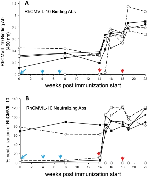
Figure 7. Stimulation of RhCMVIL-10 binding and neutralizing antibodies (Ab) following immunization of RhCMV-infected rhesus macaques.
(A) RhCMVIL-10-binding antibodies were analyzed throughout the immunization schedule (Table 2) by ELISA measured at absorbance at 450 nm (see Methods for details). (B) Neutralization of RhCMVIL-10 WT biological activity by plasma collected throughout the immunization schedule (Table 2). Percent (%) neutralization of RhCMVIL-10 denotes the ratio ([ IL-12 ] Plasma+RhCMVIL-10 / [ IL-12 ]Plasma )*100, such that 100% corresponds to complete inhibition of RhCMVIL-10 and 0% is no inhibition. Values greater than 100 reflect errors/variations in the measured levels of IL-12 in the two samples. The times of DNA (cyan) and protein (red) vaccination are shown on the Figure with arrows. Three animals (A1–A3) were immunized with M1 (solid lines), and three animals (A4–A6) were immunized with M2 (dashed lines). The times of blood draws are noted by the solid symbols for M1 immunized animals and open symbols for M2 immunized animals. Where immunization and blood draws were performed on the same day, blood was taken prior to immunization. The shape of the symbols denotes different animals, with M1 immunized animals A1–A3 solid diamond, square, and circle, respectively. M2 immunized animals A4–A6 are denoted by open square, diamond, and circle, respectively. The same designations are used for panels A and B.

