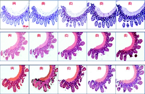
Figure 1.
Low magnification (10×) images of H&E-stained tissue histology slides for different hematoxylin and eosin staining levels. Each panel is stained with one level of eosin and five different levels of hematoxylin: (a) no eosin, (b) light eosin, and (c) normal eosin. For each panel, the five hematoxylin staining levels are: (A) very light, (B) light, (C) normal, (D) dark, and (E) very dark.

