
Fig. 4.
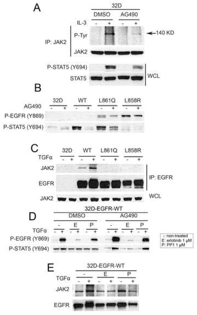
Wild-type EGFR associates with JAK2. (A) Parental 32D cells were switched for 2 h to serum-free medium containing DMSO (control) or AG490 (100 μM) and then treated with medium containing IL-3. Cell lysates were prepared and precipitated with a JAK2 antibody followed by SDS-PAGE and western blot analysis. Phospho-tyrosine band with molecular weight 140 kilodalton was detected using 4G10 antibody. Bottom two rows represent western blot analysis of whole cell lysates (WCL) using Y694 P-STAT5 and total STAT antibodies. (B) Parental 32D cells and 32D-EGFRWT, 32D-EGFRL858R, and 32D-EGFRL861Q cells were switched for 2 h to serum-free medium containing DMSO (control) or AG490. Cell lysates were analyzed by western blot with Y869 P-EGFR and Y694 P-STAT5 antibodies. (C) The indicated cells were kept in serum-free medium for 2 h and then treated with TGFα (20 ng/ml) for 3 min. Cell lysates were prepared and precipitated with cetuximab followed by SDS-PAGE and western blot analysis of immune complexes with antibodies against JAK2 and EGFR. WCL, whole cell lysates probed for JAK2 protein levels by western blot. (D) 32D-EGFRWT cells were switched to serum free medium containing DMSO, AG490, erlotinib, or PP1 where indicated and then treated with TGFα (20 ng/ml) for 3 min. Cell lysates were analyzed by western blot using antibodies against Y1068 P-EGFR and Y694 P-STAT5. (E) 32D-EGFRWT cells were kept in serum-free medium containing erlotinib (1 μM) or PP1 (1 μM) for 2 h and then treated with TGFα (20 ng/ml). Cell lysates were prepared and precipitated with cetuximab followed by SDS-PAGE and western blot analysis with the indicated antibodies.