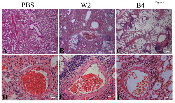
Figure 4.
Histopathology of lung tissue infected with melanin variants. Thin sections of pulmonary tissue from the uninfected mice (A, D, PBS control), or mice infected with either CBS7779-W2 (B, E) or CBS7779-B4 (C, F) (21 days post-inoculation, dpi) were stained with hematoxylin and eosin (H&E). The inflammatory response at 21 days post infection (dpi) was similar to that on 14 dpi (data not shown). Compared to the control (A), the gas exchange area in CBS7779-W2 infected lungs was largely reduced due to tissue thickening (B). Lung tissues were severely damaged in CBS7779-B4 infected mice (C). No marked inflammatory response was observed in control (D), while the infection by either CBS7779-W2 (E) or CBS7779-B4 (F) caused similar levels of peribronchial inflammation. Blood vessels within the lungs infected with CBS7779-W2 (E) were surrounded by more inflammatory infiltrates than those within CBS7779-B4 infected lungs (F). Scale bar: 100 μm in A, B, C; 10 μm in D, E, F).

