
Fig. 11.
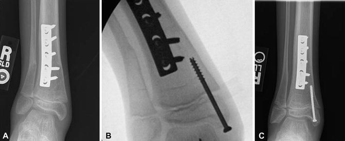
a Radiograph demonstrating distal tibia valgus deformity in a 9-year-old patient with spina bifida. b Intra-operative radiograph demonstrating the placement of a cannulated screw in the distal medial tibia. c Radiograph 19 months postoperatively demonstrating correction of the valgus alignment